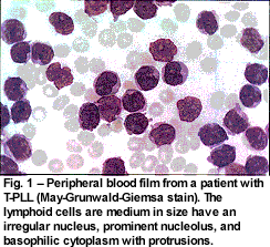
T-PLL is the condition most commonly mixed up with CLL. It comprises about 2% of mature lymphoid leukemias. It occurs in the same age group and the cells in the peripheral blood film look similar though on average the cells are slightly larger than those of CLL, the cytoplasm tends to be bluer. Although in most cases a nucleolus is visible, in 25% of cases it is not. The characteristic feature is of surface blebs in the cytoplasm. Occasionally the nucleus is very irregular.
The immunophenotype shows positivity for CD2, CD3, CD5 and CD7. B-cell markers are negative. Most cases are CD4+, CD8-; but in 15% it is the other way round and in 25% they are doubly positive. TCL1 overexpression can be demonstrated by immunohistochemistry and the TCR genes are clonally rearranged. The commonest chromosomal abnormality is inversion 14 (q11;q32) which is seen in 80% of cases and in 10% there is a reciprocal translocation t(14;14)(q11;q32). These all involve the TCA@ and TCL1A and TCL1B loci. Often the karyotype is complex with abnormalities of chromosome 8, deletions at 12p13 and 11q23 and sometimes p53 abnormalities.
T-PLL is more aggressive than CLL. It presents with an enlarged liver and spleen as well as widespread lymph node enlargement. The skin is involved in 20% of cases. Anemia and thrombocytopenia are usual and the white count usually exceeds 100. Serum immunoglobulins and HTLV1 serology are normal.
The median survival is less than a year, although more chronic cases have been reported (I saw one patient who responded well to chlorambucil for more than two years). The best responses have been seen with Campath. A trial of PARP1 inhibitors has started. Stem cell transplant should be explored in patients who are young enough and who have a donor.

The other condition most commonly confused with CLL is large granular lymphocytic leukemia (LGL leukemia). This is a heterogeneous disorder characterized by a persistent increase in large granular lymphocytes (usually between 2 and 20) without an obvious cause. LGL leukemia comprises 2-3% of mature lymphoid leukemias. Most cases are CD3+, CD8+ and show a clonal rearrangement of the TCR alpha beta genes, but occasional cases express CD4 rather than CD8 and gamma delta genes rather than alpha beta. Loss of CD5 and CD7 is rather common. Expression of CD57 and CD16 is usual. However, not all cases are derived from T-cells - some seem to be derived from NK cells and some have evidence of a mixed origin.
NK cells have CD56, CD57 and CD16 on their surface and NK-associated MHC class 1 receptors CD94/NKG2 and KIR families. Expression of a single isoform of KIR receptor is accepted as evidence of NK monoclonality. It is sometimes difficult to distinguish between T and NK LGL leukemias.
There are no characteristic chromosomal abnormalities.
Non-malignant conditions may mimic LGL leukemia. Felty's syndrom (rheumatoid arthritis with splenomegaly and neutropenia) may be a separate disease or part of the clinical picture. LGLs are increased post splenectomy, post stem cell allograft and in autoimmune diseases.
Clinically, LGL leukemias are almost always indolent. Neutropenia and moderate splenomegaly may be seen and most cases do not require treatment. Treatments that have been tried with some success include splenectomy, cyclosporin A, cyclophosphamide, steroids, low dose methotrexate and pentostatin.
 Sezary syndrome is sometimes mistaken for CLL. Although the circulating tumor cells are characteristic with 'cerebreform' nuclei, the nucleus may be contracted and quite small, so that it escapes detection. Sezary syndrome is a disseminated form of mycosis fungoides and therefore has its origin in the skin. One finds generalized erythroderma (red skin), enlarged lymph nodes and the characteristic cells in the blood.
Sezary syndrome accounts for only 5% of skin T-cell lymphomas, and since skin lymphomas are usually the province of dermatologists, they represent the only skin tumors that hematologists are likely to see.
The immunophenotype is CD2, CD3, CD5 and TCR beta positive. Most cases are CD4+ and expression of CD8 is very rare. They express the cutaneous lymphocyte antigen (CLA) and the skin-homing receptor CCR4.
The clinical features apart from those mentioned are are itching, hair loss, thickening of skin on the palms and soles, nail atrophy, and problems with the eyelids. It is an aggressive disease with only 5-10% surviving for 5 years. Treatment is experimental but may include fludarabine, pentostatin, steroids and immune therapies.
3 comments:
Nice review of a complex subject. All of the technology, even the nomenclature for the finding (FISH,flow cytometry) came out after I finished medical school. and it has been one of the pleasant challenges of having CLL to have to learn this new vocabulary and the underlying biology and technology.
If you can recommend a good overview for a family doc like myself, I would be most grateful
Thanks again for a fine post
Brian
I don't know of one, that's why I wrote this. I got my information from WHO Classification of Tumours of Haematopoietic and Lymphoid Tissues. 2008. ISBN 978-92-832-2431-0
Or did you mean a good overview of the vocabulary and technology? I don't know where that is all written down either. The book I wrote about it in 1981 is hopelessly outdated!
Terry,
I guess I am looking for a primer of understanding what the biology on the important CDs, Zap 70 and some of the critical biochemistry such as syk kinase and others.
I don't need to be an expert, but I would like some better understanding and am willing to put some effort into it.
Be well
Brian
Post a Comment